Airway fundamentals
Paediatric Upper Airway Anatomy & Development
Jump to
The whole page in one idea
Build it → separate it → open it → support it
Congenital airway problems make much more sense if you ask which developmental step went wrong. Then translate that anatomy into the problem in front of you: stridor, aspiration, a tube that will not advance, or an unexpectedly difficult extubation.[5]
01
Build it
Head, neck and laryngeal structures begin to take shape.
02
Separate it
Airway and oesophagus must become distinct pathways.
03
Open it
The laryngeal lumen must recanalise.
04
Support it
Cartilage and soft tissue must give the airway a stable lumen.
Start with normal anatomy
Why the infant airway behaves differently
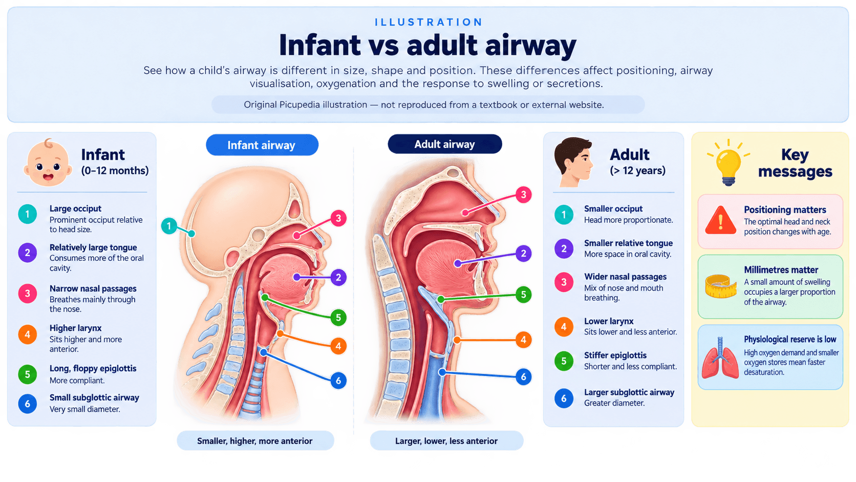
Picupedia illustration.
Selected feature
Relatively prominent occiput
What is different?
Infants have a relatively prominent occiput compared with older children and adults.[1,3]
Why does this matter?
When a small infant lies flat, the head can be pushed forwards into neck flexion, which may worsen airway alignment.
🩺 PICU pearl
Myth, memory aid… or oversimplification?
Is the paediatric larynx really a simple funnel?
Older teaching often described a uniformly funnel-shaped paediatric larynx with the cricoid as the universal narrowest point. Modern imaging shows a more complex three-dimensional airway whose dimensions vary with age, level, orientation and measurement technique.[2,4]
Embryology
The upper airway, lower airway and oesophagus do not simply appear as one finished tube. They form through coordinated growth, separation, remodelling and recanalisation. That is why a child can have a problem at one level but not another. [5]
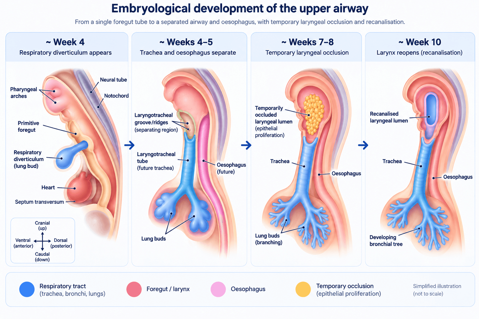
Original Picupedia teaching illustration. Simplified and not to scale.
01
Build it
Face, neck and laryngeal structures develop from multiple embryological components.
02
Separate it
The respiratory tract and oesophagus have to become distinct pathways.
03
Open it
The developing laryngeal lumen temporarily closes and then must recanalise.
04
Support it
Cartilage, muscle and connective tissue must form a stable but functional airway.
Congenital airway conditions
When development goes wrong
Congenital airway disease often reveals itself during an ordinary problem: feeding, a viral illness, intubation or extubation. The trick is to recognise when the pattern does not fit a purely functional or inflammatory explanation.
Interactive diagram
One early pathway has to become two
Normal
The respiratory tract and oesophagus become distinct channels. Air and feeds should not have a direct connection between them.
H-type TEF
The sneaky fistula
Because the oesophagus remains continuous, the child can swallow and the diagnosis may be delayed. A 2025 systematic review found bronchoscopy had the highest reported diagnostic accuracy, while contrast oesophagography could be false-negative.[7]
PICU thought
If “recurrent pneumonia” repeatedly follows feeds, do not assume the problem is only swallow coordination. Ask whether there is a structural communication.
Laryngeal cleft
Aspiration can be anatomical
A laryngeal cleft is an incomplete posterior separation between airway and oesophageal side. It ranges from a small interarytenoid defect to a much deeper cleft extending into the trachea.[8,9]
Swallow studies can demonstrate aspiration and guide feeding management, but definitive anatomical assessment generally requires specialist direct laryngoscopy/bronchoscopy with careful examination of the posterior larynx.[8,9]
Laryngeal web
A residual web can give an abnormal cry, stridor or respiratory distress. Congenital anterior glottic webs are also associated with 22q11.2 deletion, so syndromic assessment may be relevant.[19]
Subglottic stenosis
A congenitally narrow subglottic airway may first become obvious when the child develops stridor, fails extubation or needs a much smaller ETT than expected.
Laryngeal atresia
At the severe end, there is no usable laryngeal opening. Prenatal recognition matters because the airway may need to be established while placental gas exchange is still being maintained.[20]
Fetal lungs make their own fluid
During fetal life, chloride-driven secretion moves fluid into the lung lumen. That gentle distending pressure is part of normal lung growth. Near birth, the epithelium switches increasingly towards sodium-driven absorption so fluid can be cleared for air breathing.[13]
Normally
Fluid can move out through the trachea and larynx. Fetal breathing movements also move lung liquid back and forth and influence lung expansion.
CHAOS and EXIT
The placenta buys time but it does not fix the airway
Congenital high airway obstruction can produce enlarged fluid-filled lungs, dilated distal airways, diaphragmatic flattening or inversion and cardiovascular compromise. Prenatal ultrasound and fetal MRI help define the obstruction and plan delivery.[20]
Ordinary transition
Cord clamped → placental gas exchange stops → the newborn must ventilate the lungs.
EXIT principle
Placental support is deliberately maintained while a specialist team attempts to secure or create the airway before full separation from the placenta.
C or O?
Tracheal cartilage changes what the airway can tolerate
Most tracheal cartilages are C-shaped with a more flexible posterior wall. Congenital tracheal stenosis is often associated with complete circumferential rings. A fixed narrow airway has much less reserve when swelling or instrumentation is added.[10,12]
🩺 PICU pearl
Surgical concept
What is a slide tracheoplasty?
Step 1: Narrow segment
Slide tracheoplasty uses the child's own tracheal tissue to widen a stenotic segment. It is established specialist treatment for significant congenital tracheal stenosis, especially long-segment disease, but it remains major surgery requiring multidisciplinary care.[10,11,12]
This diagram shows the principle only. Real operative planning depends on the length, location, bronchial anatomy and associated cardiovascular lesions.
Evidence update
Slide tracheoplasty is established — but it is still major surgery
A 2024 systematic review of 15 studies and 845 children reported an overall postoperative mortality of 9.3%, open revision in 2.8% and endoscopic revision in 27.6%. The message is not that the operation is unsafe but that these are complex children who need experienced multidisciplinary centres and long-term airway follow-up.[11]
Apply it in real life
What should change in PICU?
The point of this page is not to make you memorise pharyngeal arches. It is to change what you notice, what you prepare and when you stop repeating the same airway manoeuvre.
Apply it in PICU
The tube passes the cords… then stops
You are intubating a 6-month-old. The laryngoscopic view is excellent. The ETT passes through the vocal cords but meets firm resistance below them at the same depth twice.
Small airway, big consequence
Why oedema matters
For ideal laminar flow, Poiseuille's relationship gives resistance as inversely proportional to radius to the fourth power:
Do not take that equation literally for every part of the upper airway because flow is often turbulent. The useful bedside principle is simply that losing a little radius in a very small airway can create a very large clinical problem.
Post-extubation stridor
Prevention starts before extubation
High-risk features include multiple or traumatic intubation attempts, a large-for-age tube, high leak pressure and known upper-airway abnormality. International paediatric ventilator-liberation guidance suggests dexamethasone at least 6 hours before extubation in children at high risk of post-extubation upper-airway obstruction, while recognising the evidence certainty is very low.[14]
Current European neonatal/infant airway guidance
What has changed from older textbook teaching?
Video laryngoscopy
Age-adapted videolaryngoscopy is recommended first choice for neonatal and infant tracheal intubation.
Apnoeic oxygenation
Recommended during neonatal intubation; consider in infants according to hypoxaemia risk and operator experience.
Limit attempts
Repeated attempts increase trauma and physiological instability. Change operator, device or strategy rather than repeating blindly.
Rescue SGA
A supraglottic airway is an important rescue oxygenation/ventilation option when intubation fails.
Cuffed tubes
Cuffed and uncuffed tubes can both be used appropriately; cuff pressure should be minimised and monitored.
Confirm placement
Use clinical assessment plus a sustained end-tidal CO2 waveform whenever applicable.
[6]
Cuffed vs uncuffed
The old “cricoid is a natural cuff” rule is too simple
Modern European guidance allows both cuffed and uncuffed tubes in neonates and infants. For cuffed tubes, use the minimum cuff pressure needed to control leak and avoid excessive mucosal pressure; the guideline gives an upper range of about 20-30 cm H2O.[6]
Down syndrome
Think multilevel airway
Children with Down syndrome can have upper-airway crowding, relative macroglossia, hypotonia, sleep-disordered breathing and smaller tracheal calibre, with subglottic stenosis or tracheobronchomalacia in some children.[17]
Airway-stop checklist
A good laryngoscopic view does not rule out a difficult airway
1
Stop
Do not force a tube through fixed resistance.
2
Oxygenate
Re-establish oxygenation and ventilation before the next plan.
3
Change something
Downsize, change device/operator/technique, or use a rescue airway as appropriate.
4
Think anatomy
Subglottic stenosis? Complete rings? Congenital tracheal stenosis? Previous airway injury?
Evidence and practice
Evidence & current practice
Common airway management has a growing trial and guideline base. Rare congenital airway disorders are different: much of the evidence comes from retrospective cohorts, systematic reviews and international expert consensus.
Practice around the world
Current European neonatal and infant airway guidance recommends an age-adapted videolaryngoscope as first choice for tracheal intubation, apnoeic oxygenation in neonates, rescue supraglottic airway use when intubation fails, and limiting repeated attempts.[6]
Resuscitation Council UK 2025 guidance similarly emphasises a difficult-airway plan, pre-oxygenation, use of familiar direct or video laryngoscopy, consideration of apnoeic oxygenation, and limiting repeated attempts while prioritising oxygenation.[15]
The APAGBI Paediatric Airway Committee is currently working on updated APAGBI/DAS difficult-intubation guidance, so older paediatric algorithms should be interpreted alongside newer evidence and local policy.[16]
Evidence update
H-type TEF: one reassuring study may not be enough
The 2025 systematic review reported bronchoscopy as the most accurate diagnostic test in the included literature, while contrast oesophagography had lower diagnostic yield and could be false-negative. Persistent symptoms still matter.[7]
Evidence update
Laryngeal cleft: management varies
60-second recap
The embryology → PICU cheat sheet
| Developmental problem | Possible result | What it may look like in PICU |
|---|---|---|
| Airway–oesophageal separation abnormal | TEF | Coughing with feeds, recurrent aspiration or lower-airway disease |
| Posterior laryngeal separation incomplete | Laryngeal cleft | Persistent aspiration despite standard feeding strategies |
| Laryngeal recanalisation incomplete | Web / stenosis / atresia | Abnormal cry, stridor, difficult airway, prenatal obstruction |
| Abnormal tracheal cartilage | Complete tracheal rings / CTS | ETT passes cords but will not advance; fixed respiratory symptoms |
| Small inflamed airway | Post-intubation oedema | Post-extubation stridor, recession, rapidly increasing work of breathing |
| Multilevel airway differences | Down syndrome airway | Obstruction or tube-size issues may occur at more than one level |
Quick check
Choose an answer and get the reasoning immediately.
Question 1
A baby has recurrent coughing with feeds and repeated lower respiratory infections. The oesophagus is patent. Which structural problem should remain on your list?
Question 2
Why can complete laryngeal obstruction produce enlarged fetal lungs?
Question 3
The ETT passes the cords but will not advance. What is the most useful next thought?
Question 4
Which statement best reflects current European neonatal/infant airway guidance?
Question 5
What is the best way to think about a child with Down syndrome and airway difficulty?
🎯 If you remember only four things
1
Separate
Airway–oesophageal separation problems can present as aspiration or recurrent respiratory disease.
2
Open
Failure of laryngeal recanalisation gives a spectrum from web to stenosis to atresia.
3
Support
Abnormal cartilage can create a fixed airway that does not tolerate swelling or a large tube.
4
Do not force it
If the ETT passes the cords but stops below them, the problem may be the anatomy, not your laryngoscopy.
Evidence
References & further reading
Priority is given to recent European/UK guidance, international consensus documents and systematic reviews, with older physiology papers retained where they remain foundational.
- 1.Di Cicco M, et al. Structural and functional development in airways throughout childhood: children are not small adults. Pediatr Pulmonol. 2021;56(1):240-251. doi:10.1002/ppul.25169
- 2.Dalal PG, et al. Pediatric laryngeal dimensions: an age-based analysis. Anesth Analg. 2009;108(5):1475-1479. doi:10.1213/ane.0b013e31819d1d99
- 3.Harless J, Ramaiah R, Bhananker SM. Pediatric airway management. Int J Crit Illn Inj Sci. 2014;4(1):65-70. doi:10.4103/2229-5151.128015
- 4.Wani TM, et al. Cricoid ring: shape, size, and variability in infants and children. Saudi J Anaesth. 2017;11(2):203-207. doi:10.4103/1658-354X.203051
- 5.Lungova V, et al. Mechanisms of larynx and vocal fold development and pathogenesis. Cell Mol Life Sci. 2020;77:3781-3795. doi:10.1007/s00018-020-03506-x
- 6.Disma N, et al. Airway management in neonates and infants: ESAIC/BJA joint guidelines. Br J Anaesth. 2024;132(1):124-144. PubMed
- 7.Anadolulu Aİ, et al. Comparison of diagnostic methods, surgical approaches and outcome for congenital H-type tracheoesophageal fistula: a systematic review. J Pediatr Surg. 2025;60(8):162343. doi:10.1016/j.jpedsurg.2025.162343
- 8.International Pediatric Otolaryngology Group. Consensus guidelines on the diagnosis and management of type I laryngeal clefts. Int J Pediatr Otorhinolaryngol. 2017. PubMed
- 9.Aldriweesh B, et al. International pediatric otolaryngology group consensus on approach to aspiration. Int J Pediatr Otorhinolaryngol. 2024;176:111810. doi:10.1016/j.ijporl.2023.111810
- 10.Sidell DR, et al. International Pediatric Otolaryngology Group consensus recommendations: evaluation and management of congenital tracheal stenosis. Int J Pediatr Otorhinolaryngol. 2022;161:111251. doi:10.1016/j.ijporl.2022.111251
- 11.Maltezeanu A, et al. Slide tracheoplasty for congenital tracheal stenosis: a systematic review. Int J Pediatr Otorhinolaryngol. 2024;182:111993. doi:10.1016/j.ijporl.2024.111993
- 12.Great Ormond Street Hospital. Tracheal stenosis / Tracheal Service. Current UK specialist service information. GOSH
- 13.Olver RE, Walters DV, Wilson SM. Developmental regulation of lung liquid transport. Annu Rev Physiol. 2004;66:77-101. doi:10.1146/annurev.physiol.66.071702.145229
- 14.Pediatric Acute Lung Injury and Sepsis Investigators (PALISI) Network. International clinical practice guidelines for pediatric ventilator liberation — executive summary. Am J Respir Crit Care Med. 2023. Guideline
- 15.Resuscitation Council UK. Paediatric Life Support Guidelines 2025. RCUK
- 16.Association of Paediatric Anaesthetists of Great Britain and Ireland. Paediatric Airway Committee — current priorities including updated APAGBI/DAS difficult intubation guidance. APAGBI
- 17.Respiratory and airway disorders in children with Down Syndrome: a review of the clinical challenges and management. 2025. PMC
- 18.van Stigt MJB, et al. International perspective of injection laryngoplasty for laryngeal cleft — a survey study. Int J Pediatr Otorhinolaryngol. 2024;186:112097. doi:10.1016/j.ijporl.2024.112097
- 19.Congenital laryngeal anomalies / laryngeal web review. PMC
- 20.Mong A, et al. Congenital high airway obstruction syndrome: MR/US findings, effect on management, and outcome. Pediatr Radiol. 2008;38:1171-1179. PubMed
Copyright note: Medical facts and concepts can be taught, but external textbook wording, figures and distinctive diagrams should not be copied. This page is independently written and the diagrams/animations above are original Picupedia teaching graphics built from the underlying clinical concepts.
Educational use only: This resource is designed for healthcare education and does not replace local guidelines, senior clinical advice, specialist airway planning or patient-specific assessment.