Case 01
A Race Against the Brain
A previously well teenager develops aphasia and dense hemiparesis immediately following a sporting injury.
The call
A previously fit and well 14-year-old is taking part in martial arts training when he sustains a blow to the left side of his head.
He stumbles forward and collapses.
Immediately afterwards, he is unable to speak and cannot move his right side.
There is no witnessed seizure.
On arrival
Pause & Decide
What's going through your mind?
The first scan
An urgent non-contrast CT head shows:
No intracranial haemorrhage.
No significant traumatic contusion is identified.
CT angiography demonstrates:
- • attenuation of the left MCA and ACA
- • more marked reduction around the proximal MCA / M1
- • mild attenuation of the distal left internal carotid artery
- • no definite intracranial thrombus
Specialist neuroradiology review considers the appearances suspicious for a left internal carotid artery dissection.
Suspected mechanism
Blunt head trauma
↓
Arterial wall injury
↓
ICA dissection / intramural haematoma
↓
Arterial narrowing ± thrombus
↓
Reduced cerebral blood flow
↓
Cerebral ischaemia
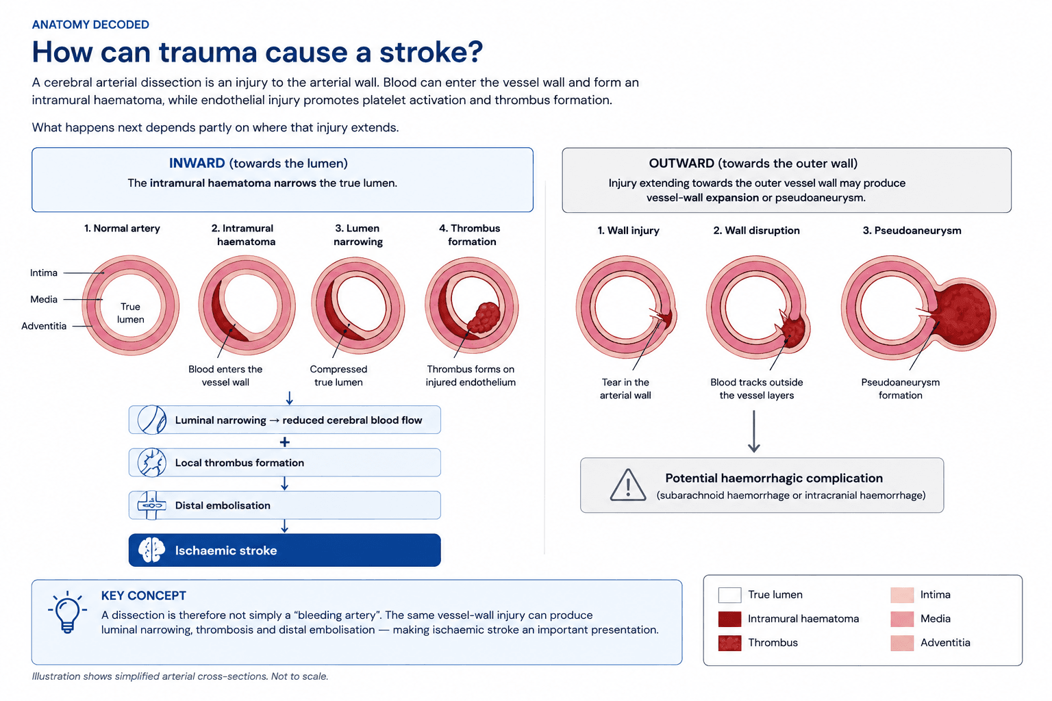
CTA decoded
What does “attenuation” actually mean?
On CT angiography, contrast normally produces bright opacification of the arterial lumen.
When a vessel is described as attenuated, it generally means that it appears reduced in calibre and/or less well opacified than expected.
Expected
Well-opacified vessel with preserved calibre
Attenuated
Reduced calibre and/or reduced contrast opacification
Possible explanations include stenosis, dissection, occlusion, vasospasm, reduced flow and technical factors related to contrast timing.
In this case, the combination of sudden focal neurological deficit, trauma, distal ICA asymmetry and reduced downstream MCA/ACA flow made arterial dissection an important diagnostic consideration.
No visible clot ≠ no ischaemic stroke.
Pause & Decide
Would you get an MRI/MRA now?
Evidence
Can trauma really cause stroke in a child?
Yes. Craniocervical arterial dissection is a recognised cause of childhood arterial ischaemic stroke, although it remains uncommon.
In a review of 182 reported paediatric craniocervical dissections, head or neck trauma was associated with approximately 56% of cases.
Hemiparesis was the most frequently reported clinical presentation.
The important clinical lesson is therefore not to allow the history of trauma to prematurely close the diagnosis.
Trauma explains the mechanism.
It does not necessarily explain the neurology.
Sudden aphasia, hemiparesis, gaze deviation or another clearly focal neurological deficit following head or neck trauma should raise concern for vascular injury and arterial ischaemic stroke, alongside traumatic brain injury.
Now the clock matters
The neurological deficit remains severe.
Reperfusion window
Closing
The PedNIHSS dilemma
Is 26 too high to thrombolyse?
A PedNIHSS of 26 tells us that this child has a very severe neurological deficit. But PedNIHSS measures clinical stroke severity — it does not measure infarct size, tissue viability or the amount of salvageable brain.
So why does the UK childhood stroke pathway stop at 24?
The RCPCH childhood stroke guideline recommends considering IV thrombolysis in selected children when the PedNIHSS is between 4 and 24, treatment can be given within 4.5 hours, haemorrhage has been excluded and appropriate vascular imaging criteria are met.
RCPCH thrombolysis criterion
PedNIHSS 4–24
Importantly, 24 should not be interpreted as a biological threshold at which thrombolysis suddenly stops working. The paediatric evidence base for thrombolysis is small, and this RCPCH recommendation was reached by Delphi consensus rather than a paediatric randomised trial demonstrating harm above a PedNIHSS of 24.
Historically, very severe strokes also raised concern because a high stroke score may accompany a large-vessel occlusion and a large established infarct, both of which can increase the risk of haemorrhagic transformation.
But the clinical score cannot tell us how much brain is already infarcted. Modern reperfusion assessment therefore combines the neurological deficit with vascular imaging, brain imaging, time from onset and the individual bleeding risk.
PedNIHSS measures deficit — not tissue viability.
A high score should make us think about the cause of such a severe deficit, particularly a large-vessel occlusion. It should not, by itself, be interpreted as evidence that reperfusion is futile.
There is no intracranial haemorrhage but the neurological deficit followed recent trauma and the vascular abnormality may represent arterial dissection.
The reperfusion clock
Why does 4.5 hours matter?
When cerebral arterial flow falls critically, not all affected brain tissue dies immediately.
Infarct core
Brain tissue that has already sustained irreversible injury.
Penumbra
Hypoperfused, functionally impaired tissue that may still be viable if blood flow is restored.
As time passes without adequate reperfusion, progressively more threatened tissue may become irreversibly infarcted.
Time is brain.
The conventional 4.5-hour intravenous thrombolysis window is not a biological cliff edge. It reflects clinical-trial evidence defining a time period in which the overall balance between reperfusion benefit and haemorrhagic risk remains favourable in appropriately selected patients.
Contemporary adult stroke pathways can use advanced imaging to identify selected patients who may benefit beyond conventional time windows. These adult data cannot simply be extrapolated to children.
The Decision
Would you thrombolyse?
Severe disabling neurological deficit. No intracranial haemorrhage. Suspected arterial dissection. Recent trauma. The reperfusion window is closing.
What happened
The case triggered urgent multidisciplinary discussion involving the local paediatric and adult stroke teams, PICU, retrieval medicine, neuroradiology and specialist stroke clinicians.
Neurosurgical intervention was not indicated because there was no intracranial haemorrhage or other surgical lesion.
No definite targetable intracranial thrombus was identified for mechanical thrombectomy.
The competing risks were therefore considered:
Don't thrombolyse
Continued cerebral ischaemia could result in a large hemispheric infarction with permanent aphasia, hemiparesis and lifelong neurological disability.
Thrombolyse
Recent trauma creates concern about haemorrhage from an occult traumatic injury or haemorrhagic transformation of the infarct.
Following multidisciplinary discussion, the balance of benefit and risk was considered to favour reperfusion treatment.
Tenecteplase 0.25 mg/kg was administered as a single IV bolus.
What does the evidence say?
Can children be thrombolysed?
Yes, in carefully selected circumstances but the evidence base is far smaller than in adult stroke.
The UK RCPCH childhood stroke guideline recommends considering thrombolysis in selected children with acute arterial ischaemic stroke when treatment can be delivered within the reperfusion window and haemorrhage has been excluded.
But there is a problem with this case
The RCPCH criteria specify a PedNIHSS of 4-24.
This patient's PedNIHSS was approximately 26.
The older UK guideline also describes demonstration of a partial or complete intracranial arterial occlusion corresponding to the deficit - whereas this case did not show a straightforward targetable intracranial thrombus.
This therefore sits outside a simple guideline algorithm.
Why tenecteplase?
Paediatric experience with tenecteplase remains extremely limited. A 2025 prospective international safety-surveillance report identified 11 children who received tenecteplase; 10 were adolescents aged 13–17 years. Eight ultimately had stroke and three were stroke mimics. No tenecteplase-related intracranial haemorrhage was reported.
These data are reassuring but far too small to establish efficacy, optimal dosing or definitive safety in childhood stroke.
The evidence is changing
The 2026 AHA/ASA acute ischaemic stroke guideline includes paediatric interventional stroke recommendations for the first time and reflects increasing acceptance of reperfusion strategies in carefully selected children.
This does not mean that adult stroke evidence can simply be applied unchanged to children. Paediatric stroke mechanisms, arteriopathies and available evidence remain different.
Systems of care
The 1 a.m. problem
The difficult question was not simply:
“Should this child receive thrombolysis?”
There was another question:
“Who makes that decision for a 14-year-old in the middle of the night?”
Expertise involved
Paediatric neurology expertise was not immediately available out of hours, so the decision required rapid coordination across several different specialties and sites.
What if this happened at 2 p.m.?
Immediate access to a dedicated paediatric stroke team could potentially bring paediatric neurology, stroke medicine, neuroradiology, neurointervention and intensive care expertise together earlier.
The objective is not simply more opinions — it is faster coordinated decision-making while the reperfusion clock is running.
The 2026 AHA/ASA acute ischaemic stroke guideline now includes paediatric recommendations for the first time, alongside dedicated Pediatric Stroke Readiness and Pediatric Stroke Alert resources.
In the UK, the RCPCH childhood stroke guideline remains the main national paediatric framework. However, its hyperacute reperfusion recommendations pre-date many recent developments in tenecteplase and paediatric thrombectomy.
Think about your own hospital:
Who activates the paediatric stroke pathway?
Who can authorise or recommend thrombolysis?
Who reviews the vascular imaging out of hours?
How quickly can you access paediatric neurology?
Where is the nearest thrombectomy-capable centre that will treat a child?
Would those answers be different at 2 p.m. and 2 a.m.?
What about thrombectomy?
The next question in a disabling arterial ischaemic stroke should be whether there is a large-vessel occlusion that can be mechanically retrieved.
In this case, specialist review did not identify a definite targetable intracranial thrombus.
But the question remains important because paediatric thrombectomy practice is changing rapidly.
Age alone should not prevent discussion with a neurointerventional stroke centre when a child has a disabling stroke and potentially accessible large-vessel occlusion.
Pause & Decide
What if an M1 thrombus had been visible?
The retrieval question
Would you intubate?
The patient's GCS is approximately 9-10.
That number understandably makes clinicians uncomfortable.
But much of the reduction in GCS is related to profound aphasia. Meanwhile, the patient is:
- • self-ventilating
- • normally oxygenated
- • haemodynamically stable
- • coughing
- • handling secretions
- • without recurrent seizures
Don't intubate a number.
Consider airway competence, neurological trajectory, physiology and transport risk rather than GCS in isolation.
Intubation also introduces induction, sedation, positive-pressure ventilation and the possibility of haemodynamic disturbance in a patient whose cerebral perfusion may already be critically dependent on collateral blood flow.
Retrieval is part of the treatment
Reperfusion therapy is only part of stroke management.
Threatened but still viable brain tissue may be dependent on marginal collateral perfusion.
Protect the penumbra.
After thrombolysis
Following thrombolysis, the patient was transferred urgently to PICU for neurological monitoring and specialist review.
Priorities included:
- • frequent neurological observations
- • serial PedNIHSS assessment
- • maintenance of normal physiology
- • monitoring for haemorrhage
- • urgent CT if neurological deterioration occurred
- • subsequent definitive neurovascular imaging
- • planning secondary stroke prevention
Antiplatelet or anticoagulant therapy requires specialist review, particularly where cervical arterial dissection is suspected.
Discuss
What would you have done?
Would you have thrombolysed?
How much should the recent trauma change your threshold?
Does suspected cervical arterial dissection alter your decision?
Would you have obtained MRI before treatment?
Should every disabling paediatric stroke be discussed with interventional neuroradiology?
What would you do if an M1 occlusion were present but thrombectomy was unavailable locally?
Would you intubate this child for retrieval?
Who owns the paediatric stroke pathway out of hours?
Take-home points
Children have strokes - even after trauma.
Head and neck trauma can cause cervical arterial dissection.
Sudden focal neurology should trigger urgent vascular imaging.
Do not delay time-critical treatment simply to obtain perfect imaging.
Paediatric thrombolysis remains an area of limited evidence and specialist decision-making.
Always consider whether thrombectomy is an option.
Aphasia can make GCS misleadingly low.
Protecting cerebral perfusion is a core part of retrieval management.
Paediatric stroke pathways need rapid access to neuroradiology, stroke and neurointerventional expertise.
References & further reading
Key guidelines and evidence used in this discussion.
UK guidance
Royal College of Paediatrics and Child Health. Stroke in childhood: clinical guideline for diagnosis, management and rehabilitation. 2017.
View RCPCH guideline →Contemporary stroke guidance
American Heart Association / American Stroke Association. 2026 Guideline for the Early Management of Patients With Acute Ischemic Stroke.
View guideline resources →Paediatric arterial dissection
Stence NV, Fenton LZ, Goldenberg NA, et al. Craniocervical arterial dissection in children: diagnosis and treatment.
Read article →Ravindra VM, et al. Craniocervical Arterial Dissection in Children: Pathophysiology and Management. 2019.
View on PubMed →Paediatric tenecteplase
Sun LR, Lee S, Lee-Eng J, et al. Tenecteplase for the Treatment of Pediatric Arterial Ischemic Stroke: A Safety Surveillance Report. Neurology. 2025;104:e210310.
View on PubMed →PedNIHSS
Beslow LA, et al. Concurrent validity and reliability of retrospective scoring of the Pediatric National Institutes of Health Stroke Scale. Stroke. 2012.
View on PubMed →Mechanical thrombectomy
Carretta LTA, et al. Safety of mechanical thrombectomy in pediatric acute ischemic stroke: a systematic review and meta-analysis. 2025.
View on PubMed →Evidence last reviewed: August 2026.
Open Discussion
Join the clinical discussion
How would you have approached this case? Share your reasoning, experience and perspective with clinicians around the world.
What would you have done differently?
Consider thrombolysis, imaging, thrombectomy, airway management, retrieval or anything else that influenced your decision.
Sign in to contribute to this discussion.
Discussion is limited to verified healthcare professionals.
Discussion
0 contributionsNo contributions yet.
Be the first clinician to share your perspective.